Bacteriophage research has seen a resurgence in recent years due to its potential for treating antibiotic-resistant bacteria. One of the critical ways to study phages is through visualization under a microscope. Microscopy techniques have come a long way in recent years, and there are many different types of microscopes that can be used to visualize phages. The type of microscope used will depend on the specific research question and the properties of the phage being studied. For example, electron microscopes provide high-resolution images of phages, but they require special preparation and handling, whereas light microscopes are less invasive and can be used to study phages in their natural environment.
In this article, we will take a closer look at the different types of microscopes that can be used for phage visualization, the techniques employed, and the importance of studying phages in this manner. We will explore the advantages and limitations of each type of microscope and the circumstances under which they are used. Additionally, we will delve into the latest advances in phage microscopy and their implications for future research.
Types of Microscopes for Phage Visualization
There are several types of microscopes that can be used to visualize bacteriophages, each with its own advantages and limitations. The most common types of microscopes used for phage visualization include:
Light Microscopy
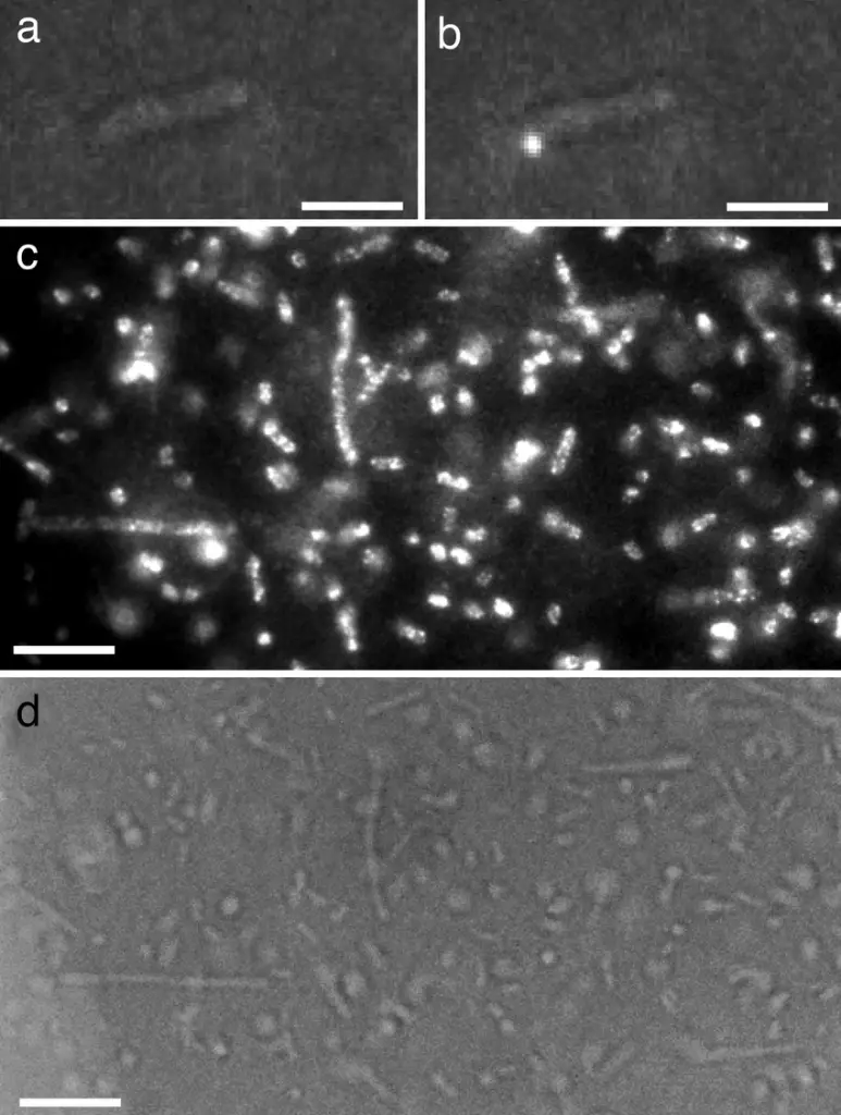
Light Microscopy: Light microscopy is the most widely used method for visualizing bacteriophages. It can be done using bright-field, phase contrast, or fluorescence techniques. Bright-field microscopy is the simplest method, where a beam of light is passed through the specimen and the image is viewed directly. This method is useful for observing the overall shape and size of the phages but may not provide the highest resolution. Phase contrast microscopy is similar to bright-field microscopy, but it uses a special condenser to enhance the contrast of the image.
Ligh microscopy can provide better (dont get it wrong, its relative) visibility of phages in solution and is commonly used for studying the behavior of phages in liquid cultures. Since most of the advanced microscopy are not well compatible with fluid environments. Scientist have tried to make stains for phages (this study used flagella stain) so that they can enhance visibility in light microscopy. Fluorescence microscopy uses a special filter to excite specific dyes in the specimen, resulting in a brightly colored image. This method is useful for identifying specific components of phages, such as viral proteins, but requires the use of fluorescent dyes and specialized equipment.
Transmission Electron Microscopy (TEM)
Transmission Electron Microscopy (TEM): TEM is a powerful technique for visualizing bacteriophages at high resolution. It involves passing a beam of electrons through a thin section of the specimen and observing the resulting image on a screen. TEM can provide images at resolutions of a few nanometers and is particularly useful for studying the structure of phages. However, this method requires the use of a vacuum chamber and can be destructive to the specimen. Additionally, samples must be prepared in a specific way, such as being embedded in resin and thinly sectioned, before they can be viewed under TEM.
Scanning Electron Microscopy (SEM)
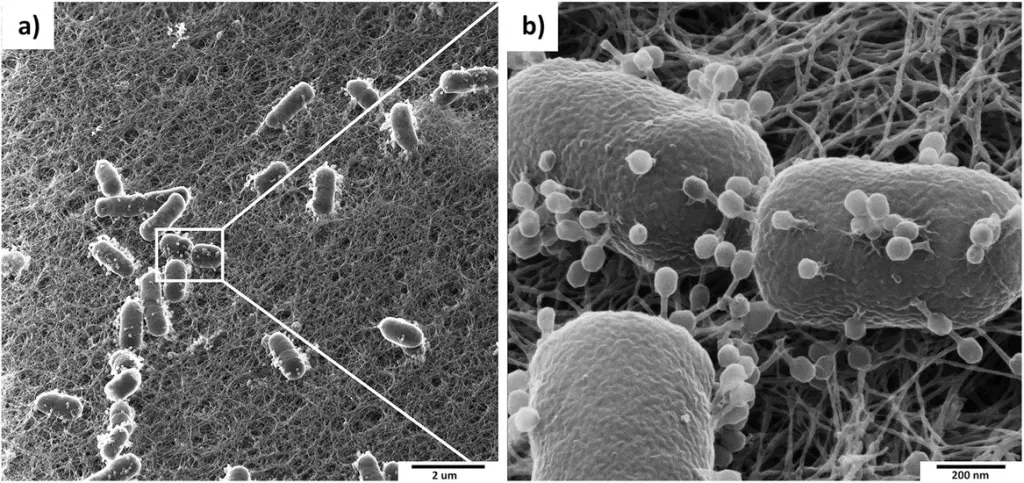
Scanning Electron Microscopy (SEM): SEM is similar to TEM but uses a beam of electrons to scan the surface of the specimen rather than passing through it. This results in images that show the surface features of the specimen in great detail. SEM is particularly useful for studying the surface morphology of phages and can be used to examine the surface features of phages in their natural state. However, this method also requires the use of a vacuum chamber and can be destructive to the specimen.
Atomic Force Microscopy (AFM)
Atomic Force Microscopy (AFM): AFM is a type of scanning probe microscopy that uses a small probe to scan the surface of the specimen. The probe is moved over the surface of the specimen, and the forces between the probe and the specimen are measured. AFM can provide images at resolutions of a few nanometers and is useful for studying the surface properties of phages. This method is particularly useful for studying the mechanical properties of phages and can be used to examine phages in their natural state without the need for a vacuum chamber. However, AFM is a relatively new technique and may not be as widely available as other methods.
Techniques for Phage Visualization
Once the type of microscope is chosen, there are various techniques that can be used to visualize phages. Some of the most commonly used techniques include:
Staining:
This involves the use of special dyes that can be used to color the phages for better visualization. Some standard staining methods include negative staining, which uses a dark background to highlight the phages, and positive staining, which uses a bright background to highlight the phages.
Labeling:
This involves the use of special proteins or other molecules that can be added to the phages to make them visible under the microscope. These include fluorescent proteins, which can be used to label phages for fluorescence microscopy, or gold nanoparticles, which can be used to label phages for electron microscopy.
Culturing:
This involves growing phages in a bacterial culture and observing them as they infect and lyse the bacteria. This technique is particularly useful for studying the life cycle of phages and their interactions with bacteria.
Phage microscopy gallery


Importance of Phage Visualization
The ability to visualize phages under a microscope is crucial for understanding their biology and potential applications. Some of the key reasons why phage visualization is important include:
- Understanding Phage Structure: By visualizing phages under a microscope, scientists can study the physical structure of these viruses in great detail. This information is crucial for understanding how phages infect and replicate within their host cells.
- Identifying Phage-Bacteria Interactions: By observing phages in their natural habitat, scientists can study how these viruses
ICTV and morphological classification of bacteriophages
In 2022, ICTV changed the classification of some bacteriophages based on their morphological taxa. Prior to that, microscopy was a valuable tool for classification. The changes impacted the morphology-based families Myoviridae, Podoviridae, and Siphoviridae, and the order Caudovirales was removed and replaced by the class Caudoviricetes to group all-tailed viruses with icosahedral capsids and double-stranded DNA genomes, both of bacterial and archaeal origin. Click here to read the full ICTV article.
The choice of a microscopy technique depends on the research question and the sample. Light microscopy is the simplest and most widely used method for visualizing bacteriophages, but it has its limitations. TEM, SEM, and AFM provide much higher-resolution images, but they are more complex and expensive. As technology advances, researchers will have access to new and more powerful tools for studying bacteriophages.
More reading
- "Phage Microscopy" by David M. White, in "Phages: Methods and Protocols" edited by Margarita Salas, Springer, 2010.
- "Phage imaging: from electron microscopy to superresolution fluorescence" by T. J. Foster and R. W. Hendrix, in "Phages: Biology and Applications" edited by R. Calendar, CRC Press, 2013.
- "Scanning electron microscopy and atomic force microscopy of bacteriophages" by J. A. McEwan, in "Phages: Methods and Protocols" edited by Margarita Salas, Springer, 2010.
- "Bacteriophage Research: A Revitalized Approach to Antibiotics" by J. Soothill, Microbiology Today, vol. 36, pp. 122-125, 2009.
- "Phage Microscopy: Techniques and Applications" by K. M. Keiler, J. Bacteriol., vol. 193, pp. 727-735, 2011.
- "Visualizing Bacteriophages Using Transmission Electron Microscopy" by L. J. Black and R. J. Doyle, Methods Enzymol., vol. 504, pp. 3-23, 2012.
- "Scanning Electron Microscopy of Bacteriophages" by P. D. R. Moineau, J. Virol. Methods, vol. 128, pp. 97-106, 2005.
- "Atomic Force Microscopy of Bacteriophages" by J. R. Parsek and E. P. Greenberg, Nat. Rev. Microbiol., vol. 2, pp. 801-811, 2004.


Comments