
Principle of Phage Plaque Assay
When a suspension of an infective phage (e.g. T4 phage) is spread over the lawn of susceptible bacterial cells (e.g. Aeromonas hydrophila), the phage attaches to the bacterial cell, replicates inside it, and kills it during its lytic release. Lysis of the bacteriophage is indicated by the formation of a zone of clearing or plaque within the lawn of bacteria. In the absence of lytic phage, the bacteria form a confluent lawn of growth.
Each plaque corresponds to the site where a single bacteriophage acted as an infectious unit and initiated its lytic cycle. The spread of infectious phage from the initially infected bacterial cell to the surrounding cells results in the lysis of the bacteria in the vicinity, eventually forming a plaque that is large enough to be visible to the naked eye. Plaques do not continue to spread indefinitely. The size of the plaque formed depends on the virus, the host, and the conditions of the culture.
The number of plaques that develop and the appropriate dilution factors can be used to calculate the number of bacteriophages i.e. plaque-forming units (PFU) in a sample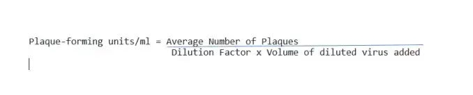
The medium used in phage plaque assays has a relatively low percentage of agar and therefore is called soft agar; it permits diffusion of phage to nearby uninfected cells but does not permit new phages to move to remote parts of the plate.
Procedure for viral (bacteriophage) Plaque
 Plaque")
- Preparation of Stock Solution by serial dilution placed eight sterile saline tubes (0.9 ml each) in your test-tube rack.
- Label one tube “control” and label the remaining five tubes consecutively from 10-1 through 10-7.
- Label six nutrient agar plates the same as the tubes.
- Using a sterile 1 ml pipette, aseptically transfer 0.1 ml of the bacteriophage suspension provided to the saline tube labeled 10-1.
- Mix the tube well by rolling it between the palms of your hands.
- With another 1 ml pipette, transfer 0.1 ml from the 10-1 tube to the 10-2 tube. Mix the tube.
- Using a fresh pipette for each transfer, transfer 0.1 ml of the suspension from the 10-2 tube to the 10-3 tube, and continue this diluting procedure consecutively for the remaining saline tubes. Do not forget to mix each tube well before and after diluting.
- Overlaying Plate with Phage-Agar Mixture
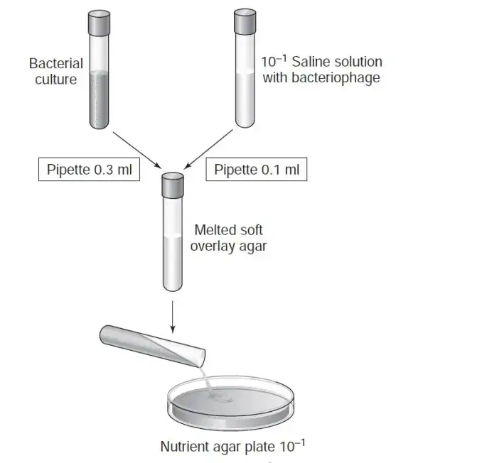
- Note: you must work quickly here) Obtain six tubes of melted soft overlay agar from the water bath. Pipette 0.3 ml of a broth culture of E.coli into each of the soft agar tubes. Mix each tube well by rolling it between your palms. Label each tube with your initials and return them to the water bath as soon as possible. Do not allow the agar to solidify.
- (Again work quickly) Remove one inoculated tube of soft agar from the water bath. Wipe off all the water from the surface of the tube. Using a 1 ml pipette, aseptically transfer 0.1 ml of the 10-1 saline phage dilution into the soft agar tube. Mix the agar tube by rolling it between your hands.
- Immediately, aseptically pour the soft agar onto the surface of the nutrient agar plate correspondingly labelled as 10-1. Replace the lid and without picking up the plate, rotate it gently in a 6-to 8-inch circle on the surface of the table to evenly distribute the agar.
- Using a fresh 1 ml pipette each time and working quickly, repeat steps 1 and 2 for the remaining saline phage dilution tubes and for the saline control tube.
- For each dilution tube, use its correspondingly labeled nutrient agar plate.
- Allow the soft agar to solidify.
- Invert and incubate plates at 35°C to 37°C for 24 hours (depend on the host in question).
There is a calculator tool we have developed that may help calculate PFU/mL after counting your plaque (PFU Calculator).


thanks for the post