The Phage

Menu

Category: MORPHOLOGY

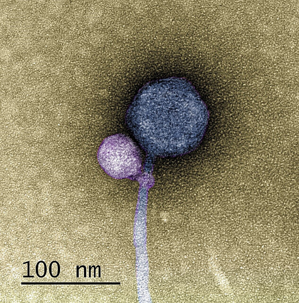
Discovery: A virus that attaches to another virus

In a groundbreaking discovery that began as a typical research project, a team from the University of Maryland, Baltimore County (UMBC) uncovered a viral phenomenon that had never been observed before: satellite viruses attaching themselves to helper viruses. This unique revelation sheds new light on the intricate relationships that exist in the phage and the whole viral world in general.

It’s a known fact that some viruses, referred to as “satellites,” rely not only on their host organism but also on another virus known as a “helper.” These helper viruses play a crucial role in either constructing the satellite’s protective shell or aiding in DNA replication. However, while it was understood that satellites and helpers had to be in proximity to each other, the actual act of attachment had never been witnessed, until now.

Published in The ISME Journal, a collaborative team from UMBC and Washington University in St. Louis reported their unprecedented observation of a satellite bacteriophage attaching itself to a helper bacteriophage at a specific point – the “neck,” where the capsid meets the tail of the virus. Astonishingly, 80 percent of the helper viruses observed had a satellite virus firmly attached at the neck, while the rest showed remnant satellite tendrils, resembling bite marks.

The team’s discovery was not just about a random interaction; it delved into the long-term relationships that exist in this viral world. By analyzing the genomes of the satellite, helper, and host, they uncovered clues about how these viruses coexist. Most satellite viruses possess a gene that allows them to integrate into the host cell’s genetic material. This integration enables the satellite to reproduce whenever a helper enters the host cell. The host cell, in turn, copies the satellite’s DNA along with its own during cell division.

However, the UMBC team discovered a satellite virus, dubbed MiniFlayer, which presented a significant departure from the norm. Unlike other satellites, MiniFlayer lacked the gene for integration. This meant that it had to remain close to its helper, named MindFlayer, every time it entered a host cell to ensure its survival. While not directly proven, this unique adaptation of attachment made sense given the circumstances.

Bioinformatics analysis further revealed that MindFlayer and MiniFlayer had co-evolved for an astonishingly long time, approximately 100 million years. This suggests that there may be numerous similar viral relationships yet to be uncovered, waiting to be explored by researchers.

The original study's TEM images showcasing First-ever observation of a virus attaching to another virus
The original study (referenced below) includes representative TEM images showcasing various scenarios: (A) MulchRoom, (B) MindFlayer, (C) MiniFlayer, (D) MiniFlayer affixed to the neck of MindFlayer, (E) MindFlayer’s neck proteins exhibiting remnants of MiniFlayer tail fibers, and (F) MindFlayer/MiniFlayer attachment to S. scabiei. Arrowheads are employed to indicate the points of connection.

The journey that led to this groundbreaking discovery was an unexpected one. It all began as a routine semester in the SEA-PHAGES program, where undergraduates isolate bacteriophages from environmental samples for analysis. However, when the University of Pittsburgh’s sequencing lab reported contamination in UMBC’s sample, the mystery began. The discovery hinged on using a transmission electron microscope at UMBC’s Keith R. Porter Imaging Facility to visualize the attachment, a tool not readily available to everyone.

This breakthrough not only raises questions about how common such attachments are but also offers a glimpse into the hidden world of viral relationships. It challenges previous assumptions about contamination and opens up new avenues for further research in the ongoing evolutionary battle between satellite and helper phages.

The UMBC team’s discovery of satellite viruses forming attachments to helper viruses has unveiled an entirely new aspect of viral interactions, challenging existing paradigms and paving the way for exciting future research read their full scientific article here: deCarvalho, T., Mascolo, E., Caruso, S.M. et al. Simultaneous entry as an adaptation to virulence in a novel satellite-helper system infecting Streptomyces species. ISME J (2023). https://doi.org/10.1038/s41396-023-01548-0

Phage Facilitates Nitrogen Metabolism in Newly Discovered Deep-sea Bacteria

Scientists have made important progress in understanding deep-sea Planctomycetes bacteria. These bacteria are found in various ecosystems and play a vital role in global environmental processes. However, studying them in their deep-sea habitat has been tough, limiting our knowledge.

In a groundbreaking study, researchers have successfully grown a new type of Planctomycetes, which they’ve named “strain ZRK32,” from sediment taken from a cold seep site on the ocean floor. They achieved this by providing the bacteria with a special mix of nutrients that included a drug called rifampicin and different forms of nitrogen. This achievement is a big step forward in our understanding of these deep-sea bacteria.

New species of marine bacteria isolated from a deep-sea cold seep
Transmission electron microscopy (TEM) was used to visualize a novel bacteria called Poriferisphaera hetertotrophicis. CM stands for “outer membrane,” Pi for “cytoplasm,” R for “ribosome,” N for “nucleoid,” ICM for “cytoplasmic membrane,” and Py for “peripla”. Credit to Rikuan Zheng

By looking at the genes, physiology, and family tree of strain ZRK32, scientists have realized it’s quite unique. They’ve given it the name “Poriferisphaera heterotrophicis” When it comes to how it grows and multiplies, this bacterium does something different from its Planctomycetes relatives; it reproduces through a process called budding.

Researchers also carried out experiments to understand what helps this bacterium grow. They discovered that giving it plenty of nutrients or adding certain nitrogen compounds, like nitrate or ammonia, made it grow better. This growth boost happens because the bacterium can make more energy through special processes called the tricarboxylic acid (TCA) cycle and the Embden–Meyerhof–Parnas (EMP) glycolysis pathway.

An interesting finding was that when strain ZRK32 was exposed to nitrate or ammonia, it continuously released a virus famously known as bacteriophages or phages. These tiny viruses didn’t harm (through bacteria cell lysis) the bacterium’s host cells, Instead, they helped the bacterium and other marine microbes use nitrogen more efficiently. This discovery shows how these viruses can change how bacteria work and provides new insights into the complicated relationships between deep-sea Planctomycetes and viruses.

In a nutshell, this research has expanded our knowledge of deep-sea Planctomycetes bacteria. It has shown how they can adapt and use nitrogen better when influenced by these special viruses. These findings help us understand the hidden world of microbes in the deep ocean.

You can read this full article on Rikuan Zheng et al, Physiological and metabolic insights into the first cultured anaerobic representative of deep-sea Planctomycetes bacteria, eLife (2023). DOI: 10.7554/eLife.89874.1 and for more news like this please visit our dedicated news page

Inoviridae (Filamentous phages): Bacteriophages with a unique replication cycle

m13 filamentous bacteriophage

Bacteriophage is a term used to describe viruses that infect bacteria and archaea. While most phages are ‘micro predators’ that kill their bacterial hosts, filamentous phages collaborate with their hosts. These viruses are even simpler than the T-phage studied by the phage group, owing to their ease of manipulating their genetic contents. Filamentous phages have a low burden on bacteria and compensate by providing services that aid in the formation of better biofilms, delivering toxins and other factors that increase virulence, or modifying their behavior to provide novel motile activity to their bacterial hosts.

Life cycle

Some aspects of the reproductive life cycle of bacteriophage M13 depicted schematically.
Some aspects of the reproductive life cycle of bacteriophage M13 are depicted schematically. Credit: what-when-how

Filamentous phage replication, like that of other bacteriophages, occurs in the cytoplasm of the host bacteria. Adsorption into the host cell occurs via pilus-mediated adsorption. Replication is governed by the ssDNA rolling circle model. DNA-templated transcription is the transcription method used. Viral extrusion is the process by which the virus exits the host cell. In Gram-negative bacteria, viral assembly occurs at the inner membrane and is mediated by a motor protein complex embedded in the membrane. This multimeric assembly complex, which includes the ATPase p1 encoded by gene 1, is thought to mediate the hydrolysis of ATP, providing energy for phage filament assembly.

Filamentous bacteriophage genome

Circular ssDNA in filamentous bacteriophages forms an antiparallel two-stranded helix (similar to A-DNA or B-DNA). Because DNA is circular, the helix ends with two loops. The packaging signal is a hairpin that targets the genome for packaging and initiates filamentous phage assembly. Phosphates along the DNA helix interact with positively charged residues of the major coat protein, resulting in the formation of a helically symmetrical tube that gives the virion its filamentous appearance. The Watson-Crick type of pairing is only maintained for about 25% of nucleotide pairs due to the lack of complementarity (with the exception of a palindromic sequence that forms a hairpin loop – the packaging signal). Some filamentous bacteriophages form a helix with phosphates in the middle and bases pointing outwards.

Structure and genome size

Filamentous phage virions are long, flexible filaments 6-7 nm in diameter and varying in length depending on the length of the packaged DNA. Multiple genomes can be assembled into long filaments that can extend up to 20 mm if the initiation or termination of filament assembly is hampered.

Physical-chemical properties

Resistance to a wide range of pH and temperatures is a common feature of filamentous phage virions. They are nonionic detergent resistant, and some are also ionic detergent resistant. Chloroform sensitivity is shared by all filamentous phages. Filamentous bacteriophage virions do not contain lipids, which is surprising given that all virion proteins are integral inner membrane proteins before assembly. Filamentous bacteriophages behave like liquid crystals at high concentrations and can be aligned in a strong magnetic field. These Pf1 phage properties have been used in nuclear magnetic resonance (NMR) structural studies of other proteins by promoting protein alignment in the magnetic field.

Assembly of the filamentous phage virions (In detail)

All virion proteins are integral membrane proteins prior to virion assembly. Four of the five virion proteins have a single transmembrane a-helix near the C terminus and a few C-terminal residues in the cytoplasm (pVIII, pIII, pVII, and pIX). Three transmembrane helices are predicted for pVI, the virion’s fifth protein. The major coat protein is directed to the membrane, resulting in Ff infection and pVIII becoming one of the cell’s dominant proteins. This is unusual because the inner membrane protein translocation complex SecYEG usually limits the number of proteins that can be incorporated into the inner membrane. pVIII employs the alternative translocon YidC may aid in overcoming the SecYEG bottleneck, allowing for massive overproduction and membrane targeting.
M13 bacteriophage particle, genome, and life cycle. Credit: Smeal et al 2017
Filamentous phage assembly is a secretion-assembly process similar to pilus assembly or toxin secretion via dedicated trans-envelope protein secretion systems.’ The packaging signal interacts with two minor proteins, pVII, and pIX, as well as the inner membrane assembly complex (via pI), to initiate assembly. The assembly process then continues by adding major coat protein pVIII subunits until the entire DNA helix is covered. Virion proteins translocate from the inner membrane into the growing phage filament, which is lipid-free, during assembly. The assembly machinery must catalyze the conversion of protein-phospholipid interactions to protein-protein interactions during the transition from membrane to virion. A strict requirement for coordinated DNA helix translocation across the inner membrane with the association of the major coat protein subunits and dissociation of ssDNA-binding protein pV adds to the complexity of filamentous phage assembly. During this process, DNA serves as an axis around which the coat protein’s helical array is assembled.
Once the DNA has been completely covered by pVIII, the introduction of two other minor polypeptides, pIII and pVI, forms the virion’s terminating cap and releases the phage from the cell. In the absence of pIII or pVI, the infected cell appears to have hundreds of pili-like structures emanating from its surface.
Ff phages have provided the majority of the information about the trans-envelope phage assembly/secretion complex. The assembly is energized at the inner cell membrane; the pI subunit of the pI/pXI complex contains a crucial adenosine triphosphate (ATP)-binding Walkermotif; additionally, it was demonstrated in a semipermeable assembly system that the assembly needs both the ATP and proton-motive force. The trans envelope assembly machinery is completed by pIV, a large outer membrane channel protein that interacts with the pI/pXI complex. pIV is a member of the secretin family, which includes the outer membrane constituents of type II and type III secretion systems, and also the type-IV pilus assembly system. Secretins are large gated channels with periplasmic domains that are extensive. Although phage assembly is a more complex process than producing proteins, the filamentous phage assembly complex, which contains only three proteins, is remarkably simple in comparison to the type II and III secretion systems, which have 15 or more different proteins components. Many lysogenic filamentous phages lack an outer membrane channel and instead rely on secretins from the host type II secretion system or type IV pilus assembly system, which ‘moonlight’ as phage secretins and presumably work with the phage-encoded pI/pXI inner membrane complex.

Applications of filamentous phages

Even though these phages are non-lytic, they can still be useful. Filamentous bacteriophages play an important role in studying biological mechanisms that extend far beyond their own life cycle due to their relative simplicity and ease of genetic manipulation. Filamentous phages are used in the biotechnology industry for a variety of purposes, including:

What do Bacteriophage Diagrams Look Like? (Morphological classification of bacteriophages)

What do bacteriophages look like?

In both academic and non-academic contexts around the world, a well-shaped particle with a clearly separated head, tail (neck, sheath, base plate, and pins), and tail fibers (very perfect body) has been used to depict bacteriophages. The shape that comes to mind when someone mentions bacteriophages is not the only shape that phages can be. T-4 bacteriophages are the source of the most well-known illustration morphology. Regardless of the fact that other bacteriophages have similar or nearly identical shapes (having slightly deviations like lacking some parts or having different body parts proportions), there are some with quite a peculiar shape. Other morphologies include “pipe/thread” like structures (e.g., Inoviridae), round (e.g., Cytoviridae), and icosahedral (e.g., Leviviridae); morphologies can be used to classify bacteriophages in the absence of other parts.

T4 bacteriophage. Photo by Omar CotsFernandez for The phage
There are various theories as to why the T-phage shape is the most commonly used to graphically represent bacteriophages. Many scientists have different theories about how it came to be that way, with some claiming it was the first bacteriophage to be discovered and visualized, others claiming it’s easier to study because of its distinct body parts, and still others claiming that a significant percentage of phages look like that, while others claim its shape resembles “predator (killing machine)” compared to other morphologies. The majority of the recommendations are based on their structure, which makes studying their mechanisms and availability easier (which comprises a large percentage of the phage population).
Scientists are revealing a lot of important information that we would have missed in previous decades as sophisticated improvements in science and technology positively impact research. Although the age of Antimicrobial Resistance (AMR) has reignited interest in bacteriophage research, many scientists around the world are working together to find antibiotic alternatives. Much information has been released/revealed during this time, and humanity is now becoming more acquainted with their close “ally” in the microscopic universe (bacteriophages). The less prioritized phage morphologies are now in the spotlight as well.

Bacteriophage classification by morphology (shapes)

The International Committee on Taxonomy of Viruses is in charge of virus (including phage) classification (ICTV). This body has proposed a morphology-based classification system. This version of classification is simpler than Bradley’s (which will be discussed later) and contains only four groups: polyhedral or cubic, filamentous or pleomorphic phages.

Classification of bacteriophages based on morphologies as per The International Committee on Taxonomy of Viruses (ICTV)

Tailed phages (equivalents to 96% of the phages discovered)

Classified as order Caudovirales, divided into three families: Myoviridae, Siphoviridae, and Podoviridae.

P1 bacteriophage. Photo by Omar CotsFernandez for The phage

Bacteriophage family Example
Myoviridae phages- with icosahedral heads, contractile tails, double-stranded DNA (dsDNA)
  • Phage T4
  • Phage P1
Siphoviridae phages -with icosahedral heads, long and non-contractile tails, double-stranded DNA (dsDNA)
  • Phage λ
  • Lactococcus phage C2
Podoviridae phages- have an icosahedral head, short tails, double-stranded DNA (dsDNA)
  • Phage T7
  • Phage P22


Polyhedral or cubic phages

-classified into Microviridae, Corticoviridae, Tectiviridae, Leviviridae, and Cystoviridae.

Bacteriophages family Example
Microviridae phages- icosahedral head, virion size 27 nm, with 12 capsomers, single-stranded DNA (ssDNA)
  • Phage φX174
Corticoviridae phages- no envelope, 63 nm in size, complex capsid, lipids, dsDNA
  • Phage PM2
Tectiviridae phages- no envelope, 60 nm, flexible lipid vesicle, pseudo-tail, dsDNA
  • Phage PRD1
Leviviridae phages- no envelope, 23 nm, poliovirus-like, ssRNA
  • Phage MS2
Cystoviridae phages-with enveloped, icosahedral head, 70-80 nm, lipids, dsRNA
  • Pseudomonas ɸ6


Filamentous phages

It-Made up of three families known as Inoviridae, Lipothrixviridae, and Rudiviridae.

Bacteriophage family Example
Inoviridae phages- no envelope, long flexible filament or short straight rods, ssDNA
  • Phage M13
Lipothrixviridae phages- enveloped, rod-shaped capsid, lipids, dsDNA
  • Phage TTV1
Rudiviridae phages- Straight uncoated rods, TMV-like, dsDNA
  • Phage SIRV-1


Pleomorphic phages

Phages containing dsDNA are classified into several families: Plasmaviridae, Fuselloviridae, Guttaviridae, Bicaudaviridae, Ampullaviridae, and Globuloviridae.

Bacteriophages family Example
Plasmaviridae phages- enveloped, 80nm, with no capsid, lipids
  • Phage MVL2
Fuselloviridae phages- enveloped, tapered capsid with short spikes end, lipids.
  • Phage SSV1
Ampullaviridae phages- enveloped, bottle-shaped virion, 230 nm in length
  • Phage ABV
Guttaviridae phages- droplet-shaped
  • Phage SNDV
Bicaudaviridae phages- Lemon-shaped virions, 120X 80 nm, long tails
  • Phage ATV

Many scientists came up with different ways of classifying phages. However, the most popular style was the one published by Bradley in 1967which resulted in six bacteriophage-based on six morphological groups, with the seventh group added later by other scientists.

Classification of bacteriophages based on morphologies as described by Bradley (1967)

Type A: Bacteriophage with hexagonal head and tail with contractile sheath

These viruses have a “tadpole shape,” meaning they have a hexagonal head, a rigid tail with contractile sheath and tail fibers dsRNA, and T-even (T2, T4, T6) phages. Most of the phages are T-shaped.

Siphoviridae phage with a long
non-contractile tail. Drawn by
Omar Cots Fernandez for
The Phage


Type B: Bacteriophage with a hexagonal head and long, flexible tail

Unlike Type-A, these phages contain a hexagonal head, but they lack a contractile sheath. Its tail is flexible and may or may not have tail fiber, such as dsDNA phages, e.g., T1 and T5 phages.

Type C: Bacteriophage with a hexagonal head and short, non-contractile tail

Type C is characterized by a hexagonal head and a tail shorter than the head. The tail lacks contractile sheath and may or may not have tail fiber, such as dsDNA phages, e.g., T3 and T7.

Type D: Bacteriophage with only hexagonal head in symmetry with large capsomere on it

Type D contains a head made up of capsomers but lacks a tail, for example, ssDNA phages (e.g., φX174). The capsomeres are subunits of the capsid, an outer covering of protein that protects the genetic material.


Type E: Bacteriophage with a simple regular hexagonal head

This type consists of a head made up of small capsomers but contains no tail, for example, ssRNA phages (e.g., F2, MS2).

Type F: Bacteriophage with no head but with long flexible filament virion

These phages are named for their filamentous shape, a worm-like chain, about 6 nm in diameter and about 1000-2000 nm long.

Type G: No detectable capsid (This group was added later after Bradley’s original study publication)

This group has a lipid-containing envelope and has no detectable capsid, for example, a dsRNA phage, MV-L2.

Thanks for visiting The Phage. if you have liked it and you don’t want to miss out, feel free to subscribe to our newsletter by clicking here

This page is subjected to frequent updates.