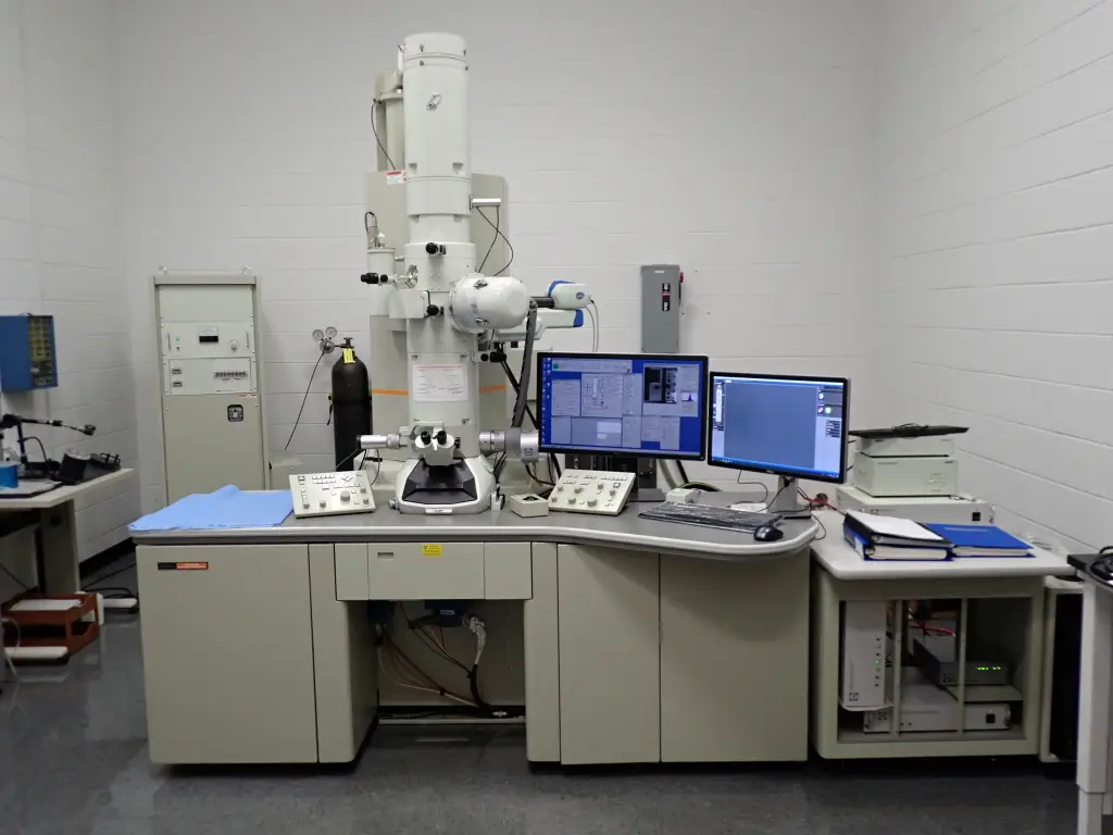
Introduction to Transmission Electron Microscopy January 17, 2024 Transmission electron microscopy (TEM) is a powerful imaging technique that uses a beam of electrons to create high-resolution images of a sample. By employing
Can next-generation sequencing replace phage microscopy? December 16, 2023 Bacteriophages, or simply phages, are viruses that infect and replicate within bacteria, playing a crucial role in shaping microbial communities and influencing bacterial evolution.
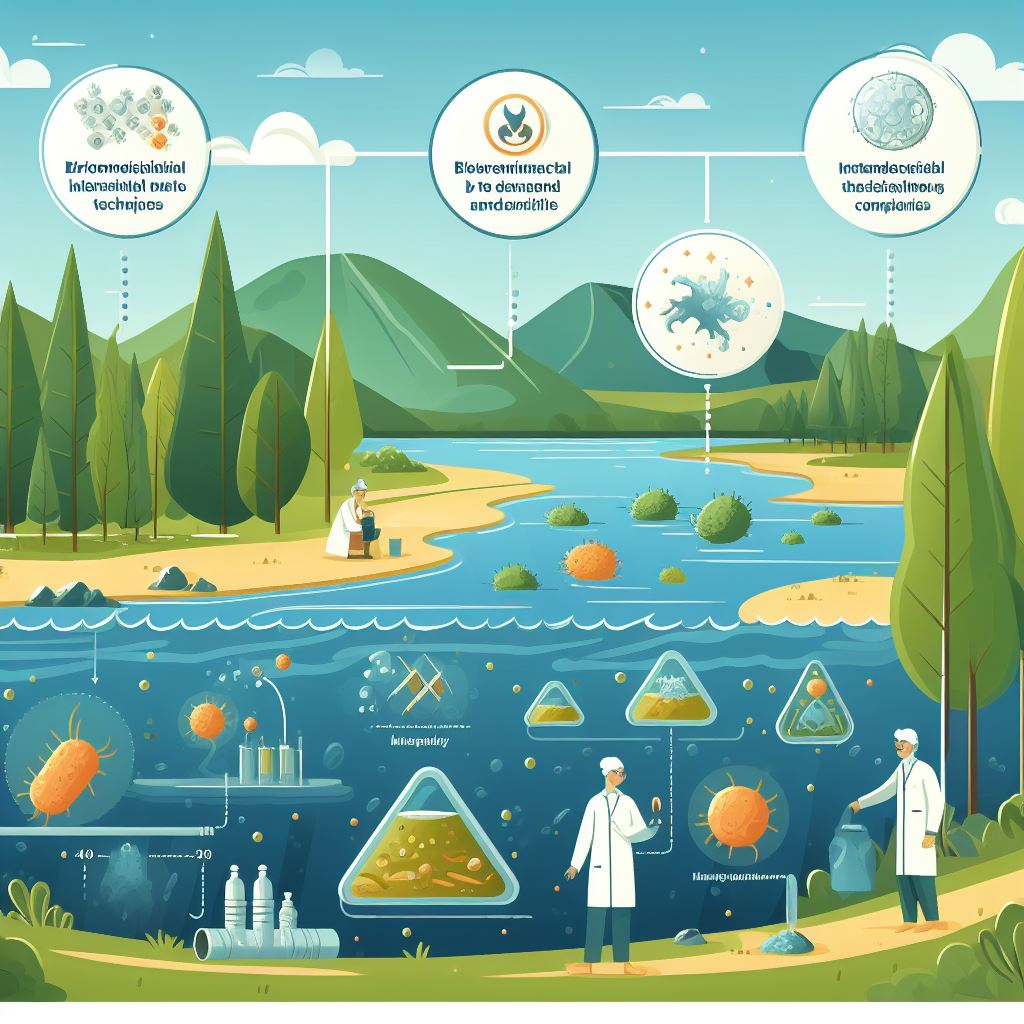
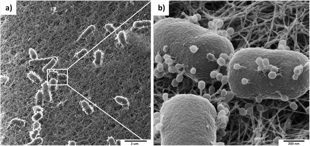
Engineered phages: The future of bioremediation? November 25, 2023 In the dynamic world of scientific discovery, researchers are constantly finding innovative solutions to some of our most pressing challenges. A recent study has
Increased Temperature Facilitates Rapid Genome Ejection in Phages November 8, 2023 In a groundbreaking study published in the Proceedings of the National Academy of Sciences in 2023, researchers from Lund University, Sweden in collaboration with
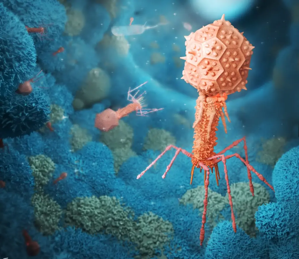
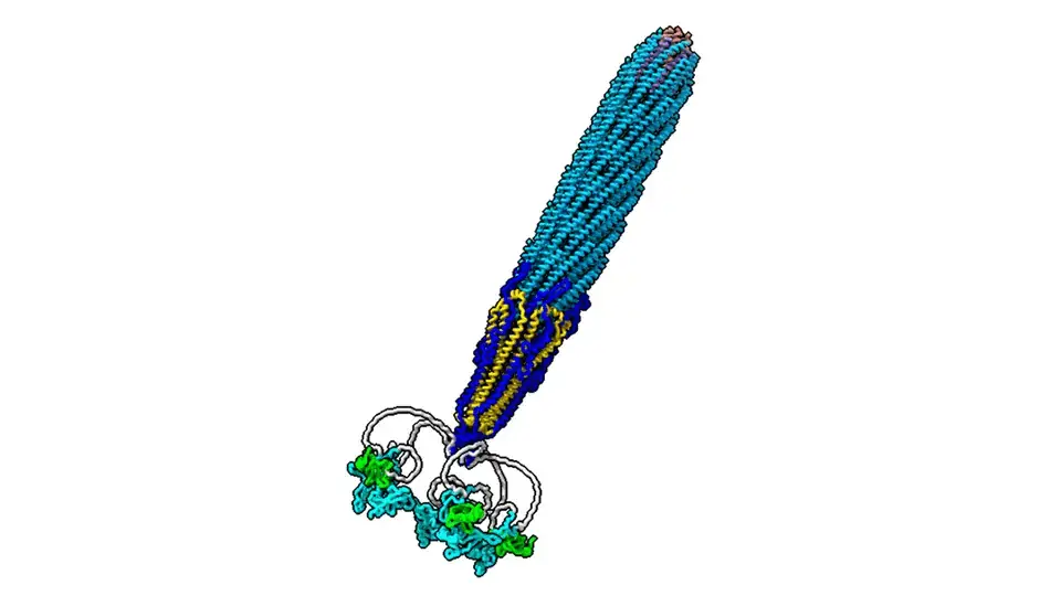
Unlocking the Secrets of Phages: Revolutionizing Biotechnology with New Structural Insights May 17, 2023 Phages, the intriguing viruses that target bacteria, have long held the key to a variety of applications in biotechnology and medicine. In a groundbreaking
Phage Visualization Under Microscope: The Types, Techniques, and Importance January 27, 2023 Bacteriophage research has seen a resurgence in recent years due to its potential for treating antibiotic-resistant bacteria. One of the critical ways to study
Problems of electron microscopy (TEM) for bacteriophages June 9, 2021 Transmission electron microscopy history Transmission electron microscope Electron microscopy has always had problems with imaging and interpretation, but the rise of digital electron microscopy